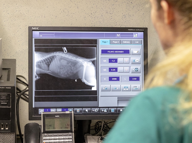
In order to provide an accurate diagnosis and the most appropriate treatment plan for your pet it is essential to have high quality diagnostic facilities.
Obtaining clear images is very important, and in order to eliminate movement blur (and to comply with Health & Safety), sedation or anaesthesia is invariably required when taking X rays.
 We have invested in the latest digital x-ray machine which produces images in remarkable detail and allows images to be viewed in both surgeries. The digital images can also be shared with other professional colleagues if needed.
While X rays are still very important there are occasions where more information is achieved with ultrasound, or with a combination of the two. Most patients tolerate ultrasound very well and allow it to be performed with them conscious.
On a less frequent basis we use endoscopes to look inside the digestive tract and on one occasion the endoscope was used to withdraw a piece of bone, which had become stuck in a dog's oesophagus/gullet. Without this the pet would have faced major chest surgery to remove it.
Smaller bronchoscopes are used to examine the throat and behind the nose, where we often find pieces of grass stuck in cats.
Our facilities also include:
(Please click on the links below to read more.)
| Consult Rooms & Pharmacy | Preparation Room | The Laboratory Facilities |
| Dentistry | Diagnostic Facilities | Hospitalisation |
| Operating Theatres |


Follow us on Facebook
4.5 Rated on Google*
* as of 14th June 2024